
1. Introduction
A choristoma is histologically normal tissue situated in an anatomically abnormal location.1 When the ectopic tissue is mature bone, the lesion is termed an osseous choristoma. Krolls et al2 used this term for mature bone in intraoral soft tissue, although older reports often referred to lingual osteoma. Vered et al3 questioned whether all such lesions share a developmental, reactive, or neoplastic origin.
Shareef et al4 identified 35 case reports or case series describing 69 patients with lingual osseous choristoma. Patients were predominantly female (48 of 69; 69.6%), with ages ranging from 5 to 73 years and a mean (SD) age of 28.1 (16.7) years. Most lesions involved the posterior third of the tongue. Of 41 lesions with recorded morphology, 33 (80.5%) were pedunculated.4
Symptoms were reported in 38 patients (55.1%); gagging or globus sensation and dysphagia were the most frequent complaints.4 We report the clinical assessment, imaging, excision, histopathological diagnosis, and follow-up of a symptomatic adolescent with a pedunculated lesion on the posterior dorsal tongue.
We structured the case according to CARE case-report principles and the SCARE 2025 guidance for surgical case reports.5,6 The literature review is focused rather than systematic and includes selected pediatric comparisons.
2. Case presentation
2.1. Patient information and clinical findings
A 15-year-old girl from Jordan was referred to the Oral and Maxillofacial Surgery Clinic at Al-Hussein Al-Salt New Hospital for evaluation and management of a posterior dorsal tongue nodule. Her complaints had lasted approximately 6 months and included mild dysphagia, intermittent nausea, and occasional pain during swallowing, particularly after hard food. The lesion was painless at the time of examination. She reported no history of trauma to the region. Medical history, dental history, and medication intake were unremarkable.
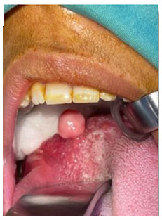
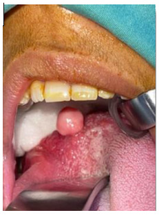
Intraoral examination showed a pink, hard, smooth, pedunculated mass covered by normal-appearing mucosa. The lesion was on the posterior dorsal tongue near the foramen cecum, slightly to the right of the midline and posterior to the circumvallate papillae. Cervical lymph-node assessment and the remaining mouth and throat examination were unremarkable. Lingual thyroid was considered in the clinical differential because of the lesion’s location. Figure 1 shows the intraoral appearance before excision.
2.2. Diagnostic assessment
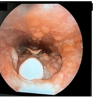
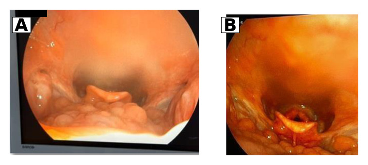
On day 1, fiberoptic nasolaryngoscopy was performed to visualize the posterior dorsal tongue. The examination showed the mass without reported airway compromise and documented its anatomical position (Figure 2).
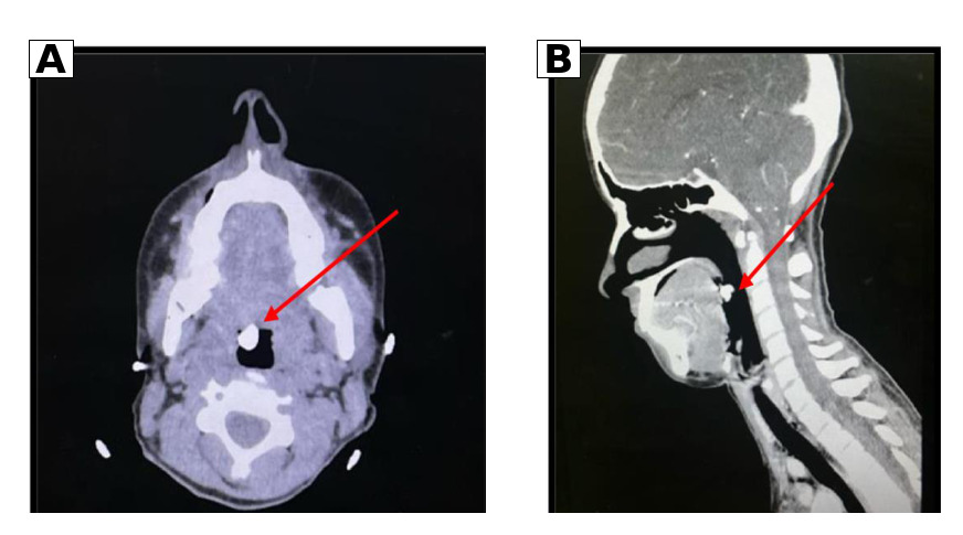
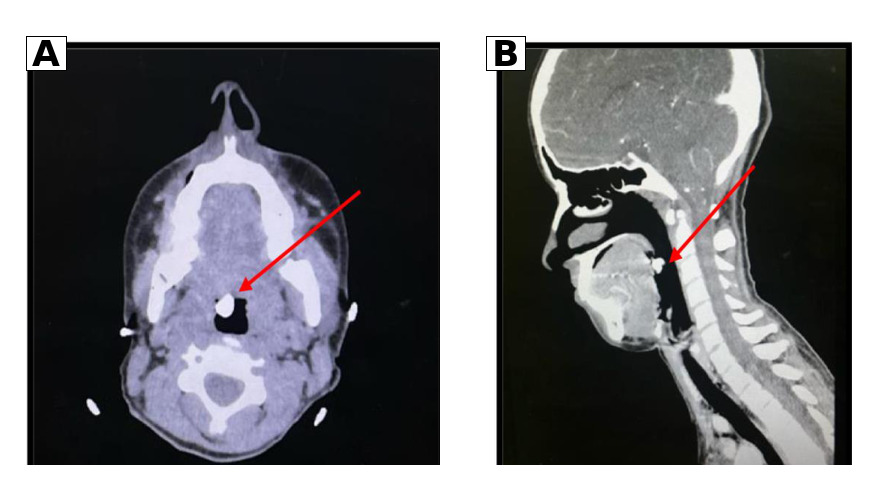
On day 4, contrast-enhanced computed tomography (CT) was performed with 1-mm slice thickness to characterize the lesion and its vascularity and to assess its anatomical relationships with adjacent structures, including the thyroid gland. Axial and sagittal images showed a hyperdense, mineralized lesion on the posterior dorsal tongue (Figure 3). The thyroid gland was normal in size and normally positioned in the neck. Imaging supported a suspected osseous choristoma, but histopathological examination was required to establish the diagnosis and distinguish it from a cartilaginous choristoma or another mineralized soft-tissue lesion.
Because the mineralized lesion was symptomatic, excisional biopsy was selected to obtain a definitive diagnosis and relieve the symptoms.
2.3. Therapeutic intervention
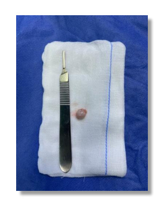
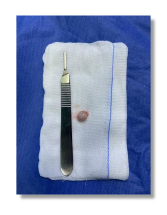
On day 15, under general anesthesia, complete local excision was performed using a No. 15 blade, and hemostasis was achieved with bipolar cautery. The specimen was submitted to the histopathology laboratory for diagnostic confirmation. The gross surgical specimen measured 0.7 × 0.6 × 0.5 cm (Figure 4).
2.4. Histopathological findings
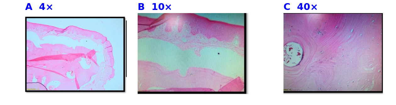
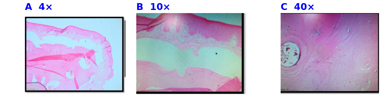
Histopathological examination showed benign surface oral mucosa with stratified squamous epithelial lining and an underlying well-defined nodule composed of lamellar bone structures surrounded by fibroconnective stroma. At higher magnification, mature bone lamellae with reactive osteocytes were seen. No cellular atypia, mitotic figures, or malignant features were identified. Histological examination confirmed clear excision margins. These findings established the diagnosis of lingual osseous choristoma (Figure 5).
2.5. Follow-up and outcomes
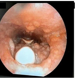
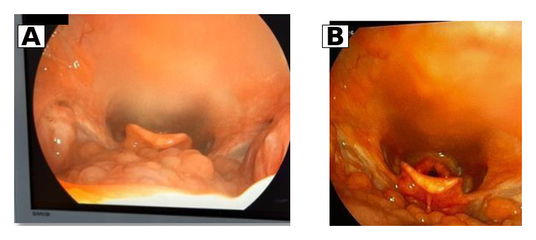
At the early postoperative review on day 19, 4 days after excision, the surgical site was healing appropriately, without surgical complications. Dysphagia, nausea, and pain during swallowing were relieved after excision and healing. Follow-up consisted of routine physical oral examinations as well as fiberoptic nasolaryngoscopy, with no clinical recurrence during four months of follow-up. No postoperative radiological imaging was performed. Postoperative endoscopic views of the excision site are shown in Figure 6.
2.6. Timeline
Exact calendar dates are withheld to protect the confidentiality of a minor.
2.7. Patient perspective
A separate patient or guardian perspective was not available because the guardians did not respond to attempts to contact them.
3. Discussion
3.1. Clinical features and pediatric comparison
The posterior dorsal location and pedunculated appearance in this patient are consistent with the commonly reported presentation of lingual osseous choristoma.4 Pediatric cases include both asymptomatic and symptomatic lesions. Arimoto et al7 and Yoshimura et al8 described asymptomatic nodules, whereas Adhikari et al9 reported a 15-year-old girl with throat discomfort and a 5-mm pedunculated lesion near the foramen cecum. Our patient presented with dysphagia, nausea, and pain during swallowing for approximately 6 months. These cases are compared in Table 1.
3.2. Pathogenesis
Proposed mechanisms include developmental tissue rests, ossification associated with thyroid or branchial arch remnants, and reactive or metaplastic ossification following chronic irritation or trauma.4,10 This patient reported no preceding trauma, but neither that history nor the presence of mature lamellar bone establishes a specific origin. The pathogenesis remains uncertain.
3.3. Differential diagnosis and diagnostic reasoning
Lingual thyroid and benign vascular or developmental lesions are relevant clinical alternatives for a nodule near the foramen cecum.8,10 In this patient, CT demonstrated mineralization and documented a normally positioned thyroid gland. The CT findings supported a suspected osseous choristoma but were not diagnostic. The distinction between osseous and cartilaginous choristoma required histopathological assessment.
The diagnosis was established by histopathological examination, which showed mature lamellar bone beneath benign stratified squamous epithelium.
3.4. Management and prognosis
Complete local excision provides tissue for diagnosis and removes the lesion. In the systematic review by Shareef et al,4 49 patients underwent surgical resection, with no recurrence reported among those cases. Our patient underwent complete local excision under general anesthesia, with histologically clear margins, uncomplicated healing, and subsequent symptom relief.
Routine physical oral examinations showed no clinical recurrence during four months of follow-up. This observation does not exclude later recurrence. Long and Koutnik11 reported recurrence in a buccal osseous choristoma; that nonlingual case does not establish a recurrence rate for lingual lesions.
3.5. Strengths and limitations
The clinical, CT, and histopathological findings document the diagnostic sequence. Interpretation is limited by the single-case design, focused literature review, and 4-month follow-up. The exact postoperative intervals for the Figure 6 photographs are unavailable, and a separate patient or guardian perspective could not be obtained. These limitations preclude conclusions about long-term recurrence or the lesion’s pathogenesis.
4. Conclusion
Osseous choristoma should be considered in the differential diagnosis of a firm, pedunculated, mineralized lesion on the posterior dorsal tongue in an adolescent. Histopathological examination established the diagnosis in this 15-year-old girl. Complete local excision with histologically clear margins was followed by healing and symptom relief, with no clinical recurrence during four months of follow-up.
Conflict of Interest / Competing Interests
The authors declare no conflicts of interest relevant to this work.
Ethics Approval and Consent
This manuscript describes a single anonymized clinical case. According to the institutional policy of the treating hospital, formal ethics committee approval was not required for publication of an anonymized case report. Consent for clinical management was obtained by the treating team. Written informed consent for publication of this case report and the accompanying images was obtained from the patient’s parent or legal guardian.
AI Tool Use Disclosure
ChatGPT (OpenAI; GPT-5) was used to assist with language editing, manuscript organization, and formatting. All clinical information, interpretations, references, and conclusions were reviewed and verified by the authors, who accept full responsibility for the final manuscript.
Third-Party Material Permissions
No third-party material requiring permission has been reproduced in this manuscript. All clinical photographs, radiographic images, histopathological images, and illustrations are original and were generated during the patient’s clinical care.
Data Availability
All data supporting the findings of this case report are included in the manuscript. No additional datasets were generated or analyzed.
Funding
This research received no external funding.
Reporting Guidelines
The report was structured according to CARE case-report principles and the SCARE 2025 guideline for surgical case reports.5,6
Preprint Disclosure
This manuscript has not been posted as a preprint and has not been previously published.
Author Contributions (CRediT)
Conceptualization: Zayed M. Al-Zu'bi (Lead). Investigation: Zayed M. Al-Zu'bi (Lead). Writing – original draft: Zayed M. Al-Zu'bi (Equal), Dareen Abu-Azzam (Equal), Esra Nsour (Equal). Methodology: Dareen Abu-Azzam (Lead). Resources: Dareen Abu-Azzam (Lead). Data curation: Esra Nsour (Lead). Validation: Esra Nsour (Lead).