
1. Introduction
Population-based studies report annual rates of 0.7 to 0.9 per 100,000 children for myocarditis-related hospitalizations in the United States and an overall incidence of 1.95 per 100,000 person-years in Finland.1,2 Despite this rarity, affected children may develop heart failure, arrhythmia, intensive care needs, and death.1 The clinical phenotype is nonspecific. In a systematic review and meta-analysis of seventeen emergency department studies comprising 737 pediatric myocarditis cases, the most common presenting features were poor feeding, fever, respiratory distress, and hypoperfusion, while elevated troponin and C-reactive protein, cardiomegaly, and ST or T-wave abnormalities were the dominant laboratory, radiographic, and electrocardiographic findings; intensive care admission was required in 88.1% of cases, and overall mortality reached 15.7%.3 In one pediatric cohort, an admission left ventricular ejection fraction below 30% was the only significant predictor of both early and late poor outcome.4
Among the viral agents implicated in pediatric myocarditis, human herpesvirus 6 (HHV-6) occupies an uncertain position. Primary infection with HHV-6B is near-universal by the age of two and usually produces exanthema subitum, a self-limited febrile illness sometimes accompanied by the rash of roseola infantum.5,6 The recognized serious complications of primary HHV-6B infection are predominantly neurological. Reviewing the disease burden of primary HHV-6B infection in immunocompetent children, Kawamura and Yoshikawa identify febrile seizures and acute encephalopathy as the most frequently reported, with primary HHV-6B infection ranking as the second most common cause of pediatric acute encephalopathy after influenza in Japan.6 Cardiac involvement during primary infection is described but uncommon. Reddy and colleagues reviewed eleven previously published cases of HHV-6-associated myocarditis or dilated cardiomyopathy in immunocompetent children and considered HHV-6 a possible causative agent, particularly in children younger than three years, while emphasizing that its pathogenic role remains uncertain because the virus is also detected in myocardium from controls.7 Causal attribution from blood PCR alone is further constrained by lifelong latency in lymphocytes and monocytes, chromosomally integrated HHV-6 in approximately 1% of the population, and the inability of qualitative blood PCR to distinguish active replication from latency or integration.5,8
This report describes the convergence of these problems in a resource-constrained center without on-site mechanical circulatory support. The patient was managed as community-acquired pneumonia, received fluid resuscitation appropriate for sepsis and dehydration but not cardiogenic shock, and progressed to refractory cardiogenic shock and death within approximately 29 hours of first presentation. A broad multiplex blood PCR panel detected HHV-6, while the other viruses included in the panel were not detected. We present the case as probable fulminant myocarditis, examine why this presentation continues to be missed, consider what an isolated qualitative blood HHV-6 result can and cannot establish about etiology, and state the diagnostic limitations directly.
2. Case Presentation
2.1. Patient Information
An 18-month-old Jordanian boy, previously healthy and developmentally normal, was brought to the emergency department on the evening of admission with a two-day history of cough and fever. The cough was dry, intermittent, and worse at night. His mother had noted increased work of breathing, described as effortful and rapid. There was no posttussive emesis, cyanosis, choking, foreign body aspiration, nasal congestion, or noisy breathing. Fever had been undocumented but intermittent and responsive to oral antipyretics. There was no rash, abnormal movement, photophobia, ear pulling, gastrointestinal symptom, change in urinary output or color, joint pain, or swelling. There had been no sick contacts, no daycare attendance, no recent travel, and no night sweats, weight loss, or loss of appetite.
His past medical and surgical history was unremarkable. He had received all immunizations on the Jordanian national schedule: BCG at birth; the hexavalent vaccine at 2, 4, and 6 months; rotavirus vaccine at 2 and 4 months; an oral polio booster at 6 months; measles vaccine at 9 months; the first measles, mumps, and rubella (MMR) vaccine and hepatitis A vaccine at 12 months; and diphtheria, tetanus, and acellular pertussis (DTaP), oral polio, and second MMR boosters at 18 months. There were no known drug or food allergies, no exposure to cigarette smoke at home, no consanguinity, and no family history of asthma, immunodeficiency, malignancy, or autoimmune disease. On directed questioning, the mother denied prior feeding intolerance, sweating with feeds, exercise intolerance, or recurrent respiratory episodes.
2.2. Clinical Findings
The patient was first assessed by the pediatric specialist in the emergency department at approximately 21:50 on the day before ward admission. He was tachypneic with effortful breathing and was described as hypoactive. He received nebulizer therapy, and the plan was admission to the pediatric ward for further management.
He arrived on the pediatric ward at 01:30. He looked unwell, tachypneic, and tachycardic. Axillary temperature was 37.0 °C, heart rate 155 beats per minute, respiratory rate 36 breaths per minute, and oxygen saturation 99% on room air; blood pressure was not recorded at the bedside on arrival. Weight was 10 kg (approximately the 10th centile), length 84 cm (approximately the 75th centile), and head circumference 46 cm (approximately the 10th centile). Nasal flaring and accessory muscle use were noted. The throat was clear, there were no palpable lymph nodes, and central cyanosis was absent. Chest expansion was symmetric, the trachea was central, percussion was resonant throughout, and air entry was good bilaterally with no added sounds. Heart auscultation revealed tachycardia. The abdomen was soft and nondistended with active bowel sounds. He was conscious and alert, with grossly intact cranial nerves, and no cerebellar signs. There was no lower limb edema or joint swelling.
The admission impression was pneumonia with respiratory distress. An electrocardiogram was not performed at admission.
2.3. Diagnostic Assessment and Investigations
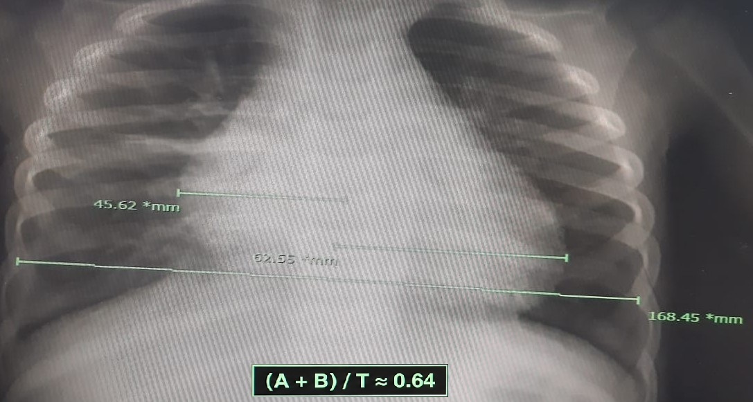
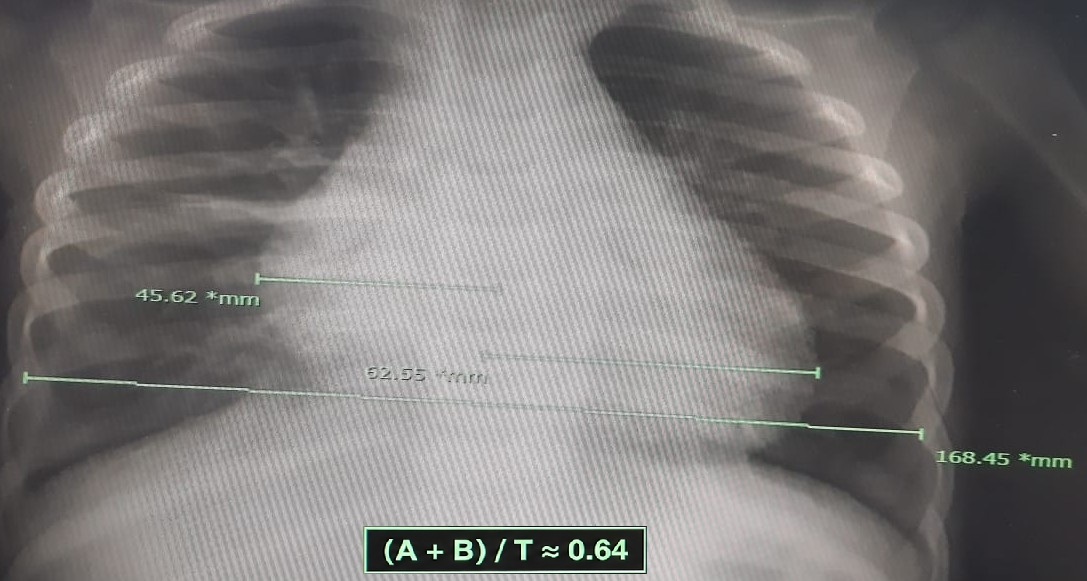
The working diagnosis at admission was community-acquired pneumonia with respiratory distress. The differential considered at the bedside on arrival to the ward included viral bronchiolitis and bacterial pneumonia with early sepsis. Cardiac etiologies were not formally entertained, given the unremarkable cardiac examination and the prominence of the respiratory presentation. Empirical management was directed at the respiratory and infectious hypotheses, with broad-spectrum antibiotics, oxygen, and scheduled nebulizers. The working diagnosis was first revised at approximately 07:00 on the day after admission, when the patient developed severe respiratory distress and hypotension that did not fit the expected pneumonia trajectory, prompting cardiology consultation and urgent echocardiography. The formal radiology report describing cardiomegaly became available at approximately 11:50. The leading clinical diagnosis became probable acute myocarditis with cardiogenic shock, while dilated cardiomyopathy remained a differential consideration.
The diagnostic challenges relevant to this case were the absence of an admission electrocardiogram, the initial bedside interpretation of the chest radiograph as compatible with pneumonia, the deferral of cardiac biomarkers and viral PCR until after clinical deterioration, and the unavailability of cardiac magnetic resonance and endomyocardial biopsy at the treating facility. At the time of cardiac evaluation, a left ventricular ejection fraction of 17%, grade 4 mitral regurgitation, and a B-type natriuretic peptide above 35,000 pg/mL collectively indicated critically severe cardiac dysfunction. The ejection fraction was below the 30% threshold associated with poor outcome in the cited pediatric cohort.4
Initial laboratory and radiographic investigations are summarized in Table 1. Relative to the reference intervals recorded by the treating laboratory, the white cell count was within range, hemoglobin was low, the platelet count was high at 604 × 103/µL on a single sample, and urea was slightly above range; creatinine was 0.37 mg/dL. Age- and assay-specific pediatric intervals were not available in the record. C-reactive protein was 5.6 times the upper reported reference limit at 2.8 mg/dL (reported range, 0 to 0.5 mg/dL).9 Blood and urine cultures were not obtained at any point during the admission. The chest radiograph obtained at admission was initially read by the on-call team as compatible with pneumonia, but the formal radiology report later that morning described cardiomegaly with bilateral perihilar haziness (Figure 1). Cardiac biomarkers, a multiplex viral PCR panel, and coagulation studies were not sent at admission; they were drawn at 13:00 on the day of deterioration. The available laboratory record identified the viral assay only as a qualitative multiplex PCR on whole blood; the platform, genomic target, HHV-6A/B differentiation, and limit of detection were not documented. SARS-CoV-2 testing was not performed, and the record did not state whether this reflected unavailability, a contemporaneous clinical judgment, or omission.
Cardiac biomarkers were measured once after deterioration. These were the first and only available values, so peak values and trends could not be determined. Repeat testing was planned for the following day, but the patient did not survive long enough for it to be performed. A capillary blood gas series across the admission captured the trajectory of metabolic compensation followed by decompensation, as shown in Table 2.
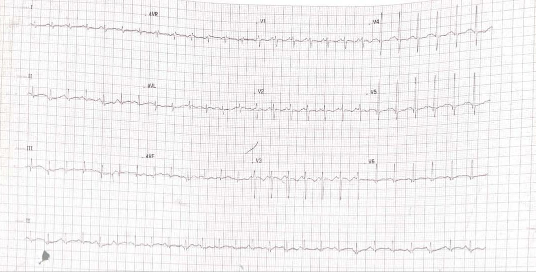
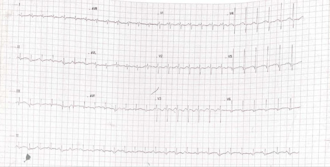
Echocardiography performed urgently after the morning deterioration showed a left ventricular ejection fraction of 17%, no pericardial effusion, grade 4 mitral regurgitation, grade 3 tricuspid regurgitation, and an estimated pulmonary artery systolic pressure of 32 mmHg. Detailed ventricular dimensions and wall thickness measurements were not formally recorded. The first electrocardiogram of the admission was obtained at the time of cardiology consultation, after the morning deterioration. The contemporaneous record described sinus tachycardia with ST deviation and T-wave abnormalities in V3 and V4 (Figure 2). The documented ST-T abnormalities contributed to the clinical suspicion of myocardial injury but do not independently confirm myocarditis.
2.4. Therapeutic Interventions and Outcomes
The clinical trajectory is summarized in Table 3.
Extracorporeal membrane oxygenation was not available at the facility, and no formal referral protocol existed for transfer to a center where it was. The family declined post-mortem examination. Because bacterial cultures, SARS-CoV-2 testing, cardiac magnetic resonance, endomyocardial biopsy, and autopsy were unavailable, the case is best categorized as probable fulminant myocarditis with qualitative blood HHV-6 detection, rather than virologically confirmed HHV-6 myocarditis.
3. Discussion
3.1. Why Fulminant Myocarditis Continues to Be Read as Pneumonia
Respiratory presentations and initial alternative diagnoses are well documented in the pediatric myocarditis literature. Rodriguez-Gonzalez and colleagues reported that 52% of pediatric myocarditis cases had received a different diagnosis at first presentation, with 31% categorized as a respiratory infection.4 The meta-analysis by Alsabri and colleagues, pooling seventeen emergency department studies, found respiratory distress in 49.8% of pediatric myocarditis cases and overall mortality of 15.7%.3 The pattern also appears in post-mortem series. In a single-center analysis of 813 pediatric autopsies over fifteen years, Neagu and colleagues identified 23 cases of histologically confirmed myocarditis, more than half in children younger than two years, and reported that myocarditis had not been suspected clinically before death.10 The American Heart Association scientific statement likewise recognizes the nonspecific presentation of pediatric myocarditis and distinguishes biopsy-proven, cardiac magnetic resonance-confirmed clinically suspected, clinically suspected, and possible disease.11
In retrospect, several features at admission warranted broader diagnostic reassessment. A heart rate of 155 beats per minute with a respiratory rate of 36 and an axillary temperature of 37.0 °C suggested disproportionate tachycardia, which in this context warranted consideration of myocardial involvement. Chest examination showed resonant percussion and good bilateral air entry without added sounds. These findings did not exclude pneumonia, but they were not strongly supportive of a lower respiratory infection severe enough to explain the observed distress. The white cell count was within the laboratory-reported interval, while C-reactive protein was elevated but nonspecific. The isolated urea and creatinine measurements were also nonspecific in this setting. None of these findings established myocarditis; their combination with persistent tachycardia and the later recognition of cardiomegaly should have widened the differential diagnosis.
A notable limitation in the initial assessment was the absence of an admission electrocardiogram. When myocarditis is suspected, electrocardiography is a standard early investigation.11,12 In the Freedman emergency department series of 31 children with myocarditis, the electrocardiogram was abnormal in 93% of cases and the chest radiograph in 55%.13 When the patient’s first electrocardiogram was obtained at cardiology consultation, more than fifteen hours after presentation, the contemporaneous record described sinus tachycardia with ST deviation and T-wave abnormalities in V3 and V4 (Figure 2). Whether an admission tracing would have shown the same abnormalities is unknown. An earlier electrocardiogram might nevertheless have widened the differential and prompted earlier echocardiography. Whether earlier recognition would have changed the outcome in the absence of mechanical circulatory support cannot be determined from this case.
The administration of two normal saline boluses totaling 35 mL/kg before probable myocarditis was recognized also illustrates the difficulty of managing undifferentiated pediatric shock. Isotonic fluid resuscitation is common when sepsis is suspected, whereas additional preload in cardiogenic shock can worsen pulmonary congestion. The 2026 Surviving Sepsis Campaign pediatric guideline recommends reassessment after each bolus and stopping further fluid administration if signs of fluid overload develop.14 At the time of the second bolus, no electrocardiogram, echocardiogram, or formal chest radiograph report was available, and pneumonia remained the working diagnosis. The case therefore illustrates how an incomplete diagnostic frame can influence fluid decisions; it does not establish that the boluses independently caused the subsequent deterioration.
3.2. HHV-6 in the Blood: What It Means and What It Does Not
Qualitative PCR on whole blood returned positive for HHV-6, while enterovirus, adenovirus, parvovirus B19, Epstein-Barr virus, cytomegalovirus, herpes simplex viruses 1 and 2, human herpesvirus 7, parechovirus, and varicella zoster virus were not detected in the same panel. These negative blood results do not exclude infection outside the panel, low-level or tissue-limited infection, or a nonviral cause. Nor does the single positive result establish HHV-6 as the myocardial pathogen.
HHV-6B seroprevalence approaches 100% by the age of two,5 and the virus establishes lifelong latency in lymphocytes and monocytes after primary infection. Detection of HHV-6 DNA in whole blood does not distinguish primary infection, reactivation, latent infection, or chromosomally integrated HHV-6, the last of which occurs in approximately 1% of the population and can produce persistently high viral DNA loads without acute disease.5,8 The Reddy review emphasizes that HHV-6 DNA can be found in heart tissue from controls and that a clinically meaningful viral-load threshold has not been established.7 Quantitative PCR with HHV-6A/B differentiation and assessment for chromosomal integration would improve interpretation. Myocardial tissue PCR, histopathology, and, where available, viral RNA assessment would provide stronger evidence of active myocardial infection, although etiologic interpretation would still require clinical correlation.5,7
Set against these caveats, the case remains reportable for three limited reasons. First, the patient was within the usual age window for primary HHV-6 infection, and rare tissue-confirmed cardiac cases have been reported in immunocompetent young children. Yoshikawa and colleagues described fatal acute myocarditis in a 5-month-old infant after roseola, with HHV-6B DNA demonstrated in myocardial tissue at autopsy.15 Grimaldi and colleagues reported a fatal case in a 23-month-old child in whom low-level HHV-6 DNA was detected in blood, while bronchoalveolar lavage also detected enterovirus and rhinovirus; their investigation did not establish a single definitive cause.16 These reports establish precedent, not attribution in the present case. Second, in an adult cohort using cardiac magnetic resonance and myocardial biopsy, HHV-6-associated myocarditis often presented with new-onset heart failure and sometimes progressed to chronic heart failure.17 Those tissue-based observations cannot be extrapolated directly to a qualitative blood PCR result in a toddler. Third, Simpson and colleagues found cardiotropic viral nucleic acid in blood more often among infants with clinical myocarditis than among controls, with HHV-6 among the viruses detected.18 This supports blood viral detection as an associated laboratory finding but not as proof of HHV-6 myocardial infection in this child.
The most defensible interpretation is that HHV-6 DNA was detected qualitatively in the blood of a child whose acute course supported probable fulminant myocarditis. Primary HHV-6 infection is biologically plausible at this age, but the negative blood results for other tested viruses do not exclude untested or tissue-limited infections. The available assay cannot distinguish primary infection, reactivation, latency, or chromosomal integration.5,7,8 Without quantitative viral load, HHV-6A/B typing, chromosomal-integration assessment, myocardial tissue PCR or viral RNA, and histopathology, the HHV-6 result cannot be assigned causal status. This case adds a hypothesis-generating observation rather than confirming HHV-6 myocarditis.
3.3. Mechanical Circulatory Support and Health-System Constraints
Mechanical circulatory support is a central rescue strategy for pediatric fulminant myocarditis with refractory cardiogenic shock. Cohort and registry studies describe the use and outcomes of extracorporeal membrane oxygenation (ECMO) and ventricular assist devices, but observational data do not by themselves establish a simple causal survival benefit.11,19,20 The American Heart Association statement nevertheless places timely mechanical circulatory support within the management pathway for the fulminant phenotype.11 Experience from lower- and middle-income settings illustrates the practical importance of access. Tuan and colleagues reported 54 pediatric patients with acute myocarditis in a Vietnamese center; 37 received ECMO, overall mortality was 30%, and median left ventricular ejection fraction at 48 hours was 25% in non-survivors and 42% in survivors.21 The present patient was managed in a facility without ECMO and without a formal referral pathway to a center where it was available. Once cardiogenic shock became refractory to inotropic and vasoactive support, no further escalation was available. The terminal capillary blood gas at 23:37 (pH 7.135, base deficit −14.3 mmol/L) documented combined respiratory and metabolic failure under the available therapy.
The intravenous immunoglobulin (IVIG) dose given was 10 g (1 g/kg), infused once during the admission. The patient died before the planned second dose. Current evidence on IVIG in pediatric myocarditis is mixed. Yen and colleagues, in a systematic review and meta-analysis of 13 studies and 1,404 children, found an unadjusted survival advantage with IVIG that lost statistical significance after correction for publication bias and concluded that routine IVIG use was not supported by the available evidence.22 A later network meta-analysis of 13 studies and 2,850 children reported associations between IVIG and lower in-hospital and overall mortality.23 The American Heart Association statement notes that IVIG is widely used but that high-quality evidence is lacking and treatment decisions should be individualized.11 This single case cannot determine whether immunomodulation altered the outcome.
A recent Jordanian case report provides a useful contextual comparison. Owdat and colleagues described a 12-month-old previously healthy girl who presented with fever, tachypnea, lethargy, and poor feeding, was diagnosed clinically with acute myocarditis, received IVIG and methylprednisolone, and survived to discharge with residual cardiac dysfunction.24 The present patient also received IVIG and methylprednisolone but did not survive. The severity of left ventricular dysfunction is not directly comparable from the published report, and numerous unmeasured differences could explain the divergent outcomes. In the Rodriguez-Gonzalez cohort, an admission ejection fraction below 30% was the only significant predictor of early and late poor outcome; the present patient’s ejection fraction was 17%.4 The comparison therefore provides regional context but does not isolate the effects of diagnostic timing, immunomodulation, or supportive care.
Reporting this case from a center without on-site ECMO provides a perspective that is less visible in literature from tertiary referral centers with full mechanical circulatory support. In many regional hospitals, a previously well child may arrive with predominantly respiratory symptoms, deteriorate rapidly, and have limited options for escalation. The clinical lesson is therefore not only to recognize a possible cardiac presentation earlier, but also to establish referral pathways before refractory shock develops.
4. Patient Perspective
Because of the fatal outcome and the family’s bereavement, a formal patient perspective could not be obtained from the patient, and the family did not provide a written statement for publication. The parents communicated to the treating team that the rapid trajectory of their previously healthy son’s illness, from a cough and fever at home to death within approximately 29 hours of first reaching medical care, was difficult to comprehend, and that they wished the clinical experience to be shared in a way that might help other families and clinicians recognize this presentation earlier. Specific reflections beyond this acknowledgment are not included, out of respect for the family’s privacy.
5. Limitations
5.1. Limitations of Clinical Management in This Case
Several aspects of the clinical management warrant acknowledgment separately from the limitations of the report. An electrocardiogram was not obtained at admission despite respiratory distress and a heart rate of 155 beats per minute in an afebrile toddler. In this setting, an electrocardiogram should have been considered as part of a broadened assessment. Cardiac biomarkers and a viral PCR panel were drawn only after deterioration, so the trajectory of myocardial injury markers from arrival to collapse is unavailable. A second normal saline bolus was administered before cardiac imaging or electrocardiography, while pneumonia remained the working diagnosis. This sequence illustrates the diagnostic risk of treating undifferentiated shock without repeated reassessment; the case cannot establish the independent effect of the fluid boluses on outcome. Blood and urine cultures were not obtained, precluding retrospective exclusion of bacterial coinfection. These observations concern the clinical sequence and its lessons, not individual blame.
5.2. Limitations of the Report
Several limitations constrain the conclusions that can be drawn from this case. No cardiac magnetic resonance or endomyocardial biopsy was performed, and no autopsy was undertaken because the family declined post-mortem examination. Cardiac histopathology, myocardial viral testing, and definitive cause-of-death attribution are therefore unavailable. The HHV-6 result was a qualitative whole-blood PCR result rather than quantitative testing; the platform, target, subtype, limit of detection, and chromosomal-integration status were not documented. SARS-CoV-2 testing was not performed, and the reason was not recorded, so COVID-19-related myocarditis or a multisystem inflammatory syndrome spectrum cannot be formally assessed. Cardiac biomarkers and the viral panel were obtained once after deterioration rather than serially or at admission, so peak values and trends cannot be reported. Blood and urine cultures were not obtained, so bacterial coinfection cannot be excluded. Echocardiographic ventricular dimensions and wall thickness measurements were not formally recorded. No admission electrocardiogram was performed, and the retained later tracing lacks documented calibration and a signed pediatric cardiologist interpretation. Finally, a single case report cannot establish causation or quantify the effect of earlier diagnosis, fluid administration, immunomodulation, or access to mechanical support.
6. Conclusion
A previously well 18-month-old boy died approximately 29 hours after first presenting with cough and fever, after a clinical course supporting probable fulminant myocarditis with qualitative blood HHV-6 detection. Cardiac magnetic resonance, endomyocardial biopsy, myocardial viral testing, and autopsy were not performed; HHV-6 myocarditis was therefore not confirmed. The practical lesson is to reconsider an exclusively respiratory diagnosis when a child treated for severe pneumonia has cardiomegaly, tachycardia disproportionate to fever, poor perfusion, hepatomegaly, or an inadequate response to respiratory therapy. These findings should prompt cardiac evaluation, including electrocardiography and echocardiography, when clinically feasible. Reassessment before additional fluid boluses may reduce the risk of worsening congestion when cardiogenic shock remains possible, and regional referral pathways for mechanical circulatory support should be established before such emergencies occur. The isolated qualitative HHV-6 result is an associated, hypothesis-generating finding, not proof of myocardial infection or viral causation.
Conflict of Interest / Competing Interests
The authors declare no conflicts of interest relevant to this work.
Ethics Approval and Consent
Written informed consent was obtained from the patient’s legal guardian for publication of this case report and the accompanying de-identified clinical data and images. Identifying details have been removed or generalized to protect patient confidentiality.
AI Tool Use Disclosure
A generative artificial intelligence tool was used to assist with language editing and editorial revision. It was not used to generate patient data or make clinical decisions. The authors reviewed and verified all clinical content, interpretations, citations, and references and take full responsibility for the final manuscript.
Third-Party Material Permissions
All tables are original. The radiograph and electrocardiogram are de-identified clinical images from the patient’s medical record. No third-party material requiring separate permission has been used.
Author Contributions (CRediT)
Conceptualization: Amro Alhyari (Supporting). Writing – original draft: Amro Alhyari (Lead). Investigation: Amro Alhyari (Lead). Data curation: Yousef H. Al Tarawneh (Lead). Formal Analysis: Yousef H. Al Tarawneh (Supporting), Bayan Maqableh (Supporting). Writing – review & editing: Yousef H. Al Tarawneh (Supporting), Bayan Maqableh (Lead). Resources: Bayan Maqableh (Supporting).
Data Availability
No research dataset was generated or analyzed for this case report. All de-identified clinical information relevant to the case is included in the article. Additional patient-level records are not publicly available because of patient confidentiality.
Funding
No funding was received for the preparation of this manuscript.
Reporting Guidelines
This case report was prepared in accordance with the CARE guidelines for case reports.
Preprint Disclosure
This manuscript has not been posted as a preprint and has not been previously published.
Abbreviations
AP, anteroposterior; BCG, Bacillus Calmette–Guérin; BNP, B-type natriuretic peptide; CARE, CAse REport; CK-MB, creatine kinase-MB; COVID-19, coronavirus disease 2019; DNA, deoxyribonucleic acid; DTaP, diphtheria, tetanus, and acellular pertussis; ECMO, extracorporeal membrane oxygenation; ESR, erythrocyte sedimentation rate; FiO₂, fraction of inspired oxygen; HCO₃-, bicarbonate; HHV-6, human herpesvirus 6; HHV-6A, human herpesvirus 6A; HHV-6B, human herpesvirus 6B; ICU, intensive care unit; IVIG, intravenous immunoglobulin; MMR, measles, mumps, and rubella; PA, posteroanterior; pCO₂, partial pressure of carbon dioxide; PC, pressure control; PCR, polymerase chain reaction; PEEP, positive end-expiratory pressure; RNA, ribonucleic acid; SARS-CoV-2, severe acute respiratory syndrome coronavirus 2; ST–T, ST-segment and T-wave.